Questo prodotto usufruisce delle SPEDIZIONI GRATIS
selezionando l'opzione Corriere Veloce in fase di ordine.
Pagabile anche con Carta della cultura giovani e del merito, 18App Bonus Cultura e Carta del Docente
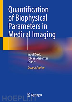
The second edition of this book offers six new chapters covering the latest developments in quantitative medical imaging, including artificial intelligence, MRI mapping, sonography, elastography and cardiac CT. All the other existing chapters have been updated and expanded, many with new text and figures, to reflect the rapid translation and advancement of technology in this exciting area of biomedical research.
This updated edition presents fundamental knowledge on the imaging quantification of biophysical parameters for clinical diagnostic purposes. Clinical imaging scanners are considered by the authors as physical measurement systems capable of quantifying intrinsic parameters for the representation of the constitution and biophysical properties of tissues in vivo. In one respect, this approach fosters the development of new imaging methods for highly reproducible, system-independent, and quantitative biomarkers. These methods are greatly detailed in the book. Alternatively, this new edition equips the reader with a better understanding of how the physical properties of tissues interact with signal generation in medical imaging, opening up new insights into the complex and fascinating relationship between structure and function in living tissues.
This updated edition is of interest to all those who recognize the limitations of clinical diagnosis based primarily on visual inspection of images, and who wish to learn more about the diagnostic potential of quantitative, biophysically-based medical imaging markers, as well as the challenges posed by the scarcity of such markers for next-generation imaging technologies.
1. Introduction.- Part 1. Biological and Physical Fundamentals.- 2. The fundamentals of transport in living tissues quantified by medical imaging technologies.- 3. Mathematical modeling of blood flow in the cardiovascular system.- 4. Equations of motion for biphasic tissue.- 5. Physical Properties of Single Cells and Collective Behavior.- 6. The extracellular matrix as a target for molecular and biophysical magnetic resonance imaging.- Part 2. Medical Imaging Technologies.- 7. Mathematical Methods in Medical Image Processing.- 8. Acceleration strategies for data sampling in MRI.- 9. Machine Learning for Quantitative MR Image Reconstruction.- 10. 4D flow MRI.- 11. New chapter with the tentative title: Quantitative multiparametric mapping MRI.- 12. CEST MRI.- 13. Innovative PET and SPECT Tracers.- 14. Methods and approaches in ultrasound elastography.- 15. Photoacoustic imaging: Principles and Applications.- 16. Fundamentals of X-Ray Computed Tomography: Acquisition and Reconstruction.- Part 3. Applications.- 17. Quantification of Myocardial Effective Transverse Relaxation Time with Magnetic Resonance at 7.0 Tesla for a Better Understanding of Myocardial (Patho)physiology.- 18. Extracellular-matrix-specific molecular MR imaging probes for the assessment of aortic aneurysms.- 19. Diffusion based MRI - imaging basics and clinical applications.- 20. Tumor characterization by ultrasound elastography and contrast-enhanced ultrasound.- 21. Quantitative Bone Ultrasound.- 22. New chapter with the tentative title: Mechanical imaging of the abdonimal aorta.- 23. Sensitivity of tissue shear stiffness to pressure and perfusion in health and disease.- 24. Radionuclide imaging of cerebral blood flow.- 25. Cardiac perfusion MRI.- 26. Myocardial Perfusion Assessment by 3D and 4D Computed Tomography.- 27. Roadmap on the use of artificial intelligence for imaging of vulnerable atherosclerotic plaque in coronary arteries.- 28. Clinical quantitative coronary artery stenosis and coronary atherosclerosis imaging: a Consensus Statement from the Quantitative Cardiovascular Imaging Study Group.
Ingolf Sack is a Heisenberg professor of the German Research Foundation for Experimental Radiology and Elastography at Charité-Universitätsmedizin Berlin, Germany. He received a PhD in Chemistry from Freie Universität Berlin for the development of methods in NMR spectroscopy. He then worked at the Weizmann Institute in Rehovot, Israel and at the Sunnybrook Hospital, Toronto. Since 2003 he has led an interdisciplinary team of physicists, engineers, chemists, and physicians who have pioneered pivotal developments in time-harmonic elastography of both MRI and ultrasound for many medical applications.
Tobias Schaeffter is the head of division of Medical Physics and metrological IT at the Physikalisch-Technische Bundesanstalt (PTB) in Berlin, Germany. He is also a Professor in Biomedical Imaging at TU Berlin and the Einstein Centre Digital Future. Tobias Schaeffter studied electrical engineering at TU-Berlin and did his PhD in magnetic resonance spectroscopic imaging (MRSI) under supervision of Prof. Leibfritz at University Bremen in 1996. From 1996-2006, he worked as a Principal Scientist at the Philips Research Laboratories in Hamburg, Germany, where he managed MR-research projects , their clinical evaluation and product integration. In April 2006, he took up the Philip Harris Professorship of Imaging Sciences at King’s College London. In 2012 he became department head of biomedical engineering. Since 2015 he moved to PTB as head of division. A major aim of his research is the investigation of fast and quantitative MR-techniques for cardiovascular applications.


Il sito utilizza cookie ed altri strumenti di tracciamento che raccolgono informazioni dal dispositivo dell’utente. Oltre ai cookie tecnici ed analitici aggregati, strettamente necessari per il funzionamento di questo sito web, previo consenso dell’utente possono essere installati cookie di profilazione e marketing e cookie dei social media. Cliccando su “Accetto tutti i cookie” saranno attivate tutte le categorie di cookie. Per accettare solo deterninate categorie di cookie, cliccare invece su “Impostazioni cookie”. Chiudendo il banner o continuando a navigare saranno installati solo cookie tecnici. Per maggiori dettagli, consultare la Cookie Policy.