Questo prodotto usufruisce delle SPEDIZIONI GRATIS
selezionando l'opzione Corriere Veloce in fase di ordine.
Pagabile anche con Carta della cultura giovani e del merito, 18App Bonus Cultura e Carta del Docente
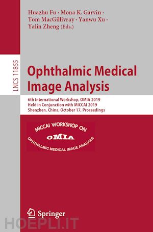
This book constitutes the refereed proceedings of the 6th International Workshop on Ophthalmic Medical Image Analysis, OMIA 2019, held in conjunction with the 22nd International Conference on Medical Imaging and Computer-Assisted Intervention, MICCAI 2019, in Shenzhen, China, in October 2019.
The 22 full papers (out of 36 submissions) presented at OMIA 2019 were carefully reviewed and selected. The papers cover various topics in the field of ophthalmic image analysis.
Dictionary Learning Informed Deep Neural Network with Application to OCT Images.- Structure-aware Noise Reduction Generative Adversarial Network for Optical Coherence Tomography Image.- Region-Based Segmentation of Capillary Density in Optical Coherence Tomography Angiography.- An ampli?ed-target loss approach for photoreceptor layer segmentation in pathological OCT scans.- Foveal avascular zone segmentation in clinical routine ?uorescein angiographies using multitask learning.- Guided M-Net for High-resolution Biomedical Image Segmentation with Weak Boundaries.- 3D-CNN for Glaucoma Detection using Optical Coherence Tomography.- Semi-supervised Adversarial Learning for Diabetic Retinopathy Screening.- Shape Decomposition of Foveal Pit Morphology using Scan Geometry Corrected OCT.- U-Net with spatial pyramid pooling for drusen segmentation in optical coherence tomography.- Deriving Visual Cues from Deep Learning to Achieve Subpixel Cell Segmentation in Adaptive Optics Retinal Images.- Robust Optic Disc Localization by Large Scale Learning.- The Channel Attention based Context Encoder Network for Inner Limiting Membrane Detections.- Fundus Image based Retinal Vessel Segmentation Utilizing A Fast and Accurate Fully Convolutional Network.- Network pruning for OCT image classi?cation.- An improved MPB-CNN segmentation method for edema area and neurosensory retinal detachment in SD-OCT images.- Encoder-Decoder Attention Network for Lesion Segmentation of Diabetic Retinopathy.- Multi-Discriminator Generative Adversarial Networks for improved thin retinal vessel segmentation.- Fovea Localization in Fundus Photographs by Faster R-CNN with Physiological Prior.- Aggressive Posterior Retinopathy of Prematurity Automated Diagnosis via a Deep Convolutional Network.- Automated Stage Analysis of Retinopathy of Prematurity Using Joint Segmentation and Multi-Instance Learning.- Retinopathy Diagnosis using Semi-supervised Multi-channel Generative Adversarial Network.


Il sito utilizza cookie ed altri strumenti di tracciamento che raccolgono informazioni dal dispositivo dell’utente. Oltre ai cookie tecnici ed analitici aggregati, strettamente necessari per il funzionamento di questo sito web, previo consenso dell’utente possono essere installati cookie di profilazione e marketing e cookie dei social media. Cliccando su “Accetto tutti i cookie” saranno attivate tutte le categorie di cookie. Per accettare solo deterninate categorie di cookie, cliccare invece su “Impostazioni cookie”. Chiudendo il banner o continuando a navigare saranno installati solo cookie tecnici. Per maggiori dettagli, consultare la Cookie Policy.