Questo prodotto usufruisce delle SPEDIZIONI GRATIS
selezionando l'opzione Corriere Veloce in fase di ordine.
Pagabile anche con Carta della cultura giovani e del merito, 18App Bonus Cultura e Carta del Docente
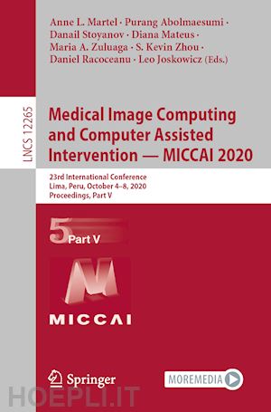
The seven-volume set LNCS 12261, 12262, 12263, 12264, 12265, 12266, and 12267 constitutes the refereed proceedings of the 23rd International Conference on Medical Image Computing and Computer-Assisted Intervention, MICCAI 2020, held in Lima, Peru, in October 2020. The conference was held virtually due to the COVID-19 pandemic.
The 542 revised full papers presented were carefully reviewed and selected from 1809 submissions in a double-blind review process. The papers are organized in the following topical sections:
Part I: machine learning methodologies
Part II: image reconstruction; prediction and diagnosis; cross-domain methods and reconstruction; domain adaptation; machine learning applications; generative adversarial networks
Part III: CAI applications; image registration; instrumentation and surgical phase detection; navigation and visualization; ultrasound imaging; video image analysis
Part IV: segmentation; shape models and landmark detection
Part V: biological, optical, microscopic imaging; cell segmentation and stain normalization; histopathology image analysis; opthalmology
Part VI: angiography and vessel analysis; breast imaging; colonoscopy; dermatology; fetal imaging; heart and lung imaging; musculoskeletal imaging
Part VI: brain development and atlases; DWI and tractography; functional brain networks; neuroimaging; positron emission tomography
Biological, Optical, Microscopic Imaging.- Channel Embedding for Informative Protein Identification from Highly Multiplexed Images.- Demixing Calcium Imaging Data in C. elegans via Deformable Non-negative Matrix Factorization.- Automated Measurements of Key Morphological Features of Human Embryos for IVF.- A Novel Approach to Tongue Standardization and Feature Extraction.- Patch-based Non-Local Bayesian Networks for Blind Confocal Microscopy Denoising.- Attention-guided Quality Assessment for Automated Cryo-EM Grid Screening.- MitoEM Dataset: Large-scale 3D Mitochondria Instance Segmentation from EM Images.- Learning Guided Electron Microscopy with Active Acquisition.- Neuronal Subcompartment Classification and Merge Error Correction.- Microtubule Tracking in Electron Microscopy Volumes.- Leveraging Tools from Autonomous Navigation for Rapid, Robust Neuron Connectivity.- Statistical Atlas of C.elegans Neurons.- Probabilistic Segmentation and Labeling of C. elegans Neurons.- Segmenting Continuous but Sparsely-Labeled Structures in Super-Resolution Microscopy Using Perceptual Grouping.- DISCo: Deep learning, Instance Segmentation, and Correlations for cell segmentation in calcium imaging.- Isotropic Reconstruction of 3D EM Images with Unsupervised Degradation Learning.- Background and illumination correction for time-lapse microscopy data with correlated foreground.- Joint Spatial-Wavelet Dual-Stream Network for Super-Resolution.- Towards Neuron Segmentation from Macaque Brain Images: A Weakly Supervised Approach.- 3D Reconstruction and Segmentation of Dissection Photographs for MRI-free Neuropathology.- DistNet: Deep Tracking by displacement regression: application to bacteria growing in the Mother Machine.- A weakly supervised deep learning approach for detecting malaria and sickle cell anemia in blood films.- Imaging Scattering Characteristics of Tissue in Transmitted Microscopy.- Attention based multiple instance learning for classification of blood cell disorders.- A generative modeling approach for interpreting population-level variability in brain structure.- Processing-Aware Real-Time Rendering for Optimized Tissue Visualization in Intraoperative 4D OCT.- Cell Segmentation and Stain Normalization.- Boundary-assisted Region Proposal Networks for Nucleus Segmentation.- BCData: A Large-Scale Dataset and Benchmark for Cell Detection and Counting.- Weakly-Supervised Nucleus Segmentation Based on Point Annotations: A Coarse-to-Fine Self-Stimulated Learning Strategy.- Structure Preserving Stain Normalization of Histopathology Images Using Self Supervised Semantic Guidance.- A Novel Loss Calibration Strategy for Object Detection Networks Training on Sparsely Annotated Pathological Datasets.- Histopathological Stain Transfer Using Style Transfer Network With Adversarial Loss.- Instance-aware Self-supervised Learning for Nuclei Segmentation.- StyPath: Style-Transfer Data Augmentation For Robust Histology Image Classification.- Multimarginal Wasserstein Barycenter for Stain Normalization and Augmentation.- Corruption-Robust Enhancement of Deep Neural Networks for Classification of Peripheral Blood Smear Images.- Multi-Field of View Aggregation and Context Encoding for Single-Stage Nucleus Recognition.- Self-Supervised Nuclei Segmentation in Histopathological Images Using Attention.- FocusLiteNN: High Efficiency Focus Quality Assessment for Digital Pathology.- Histopathology Image Analysis.- Pairwise Relation Learning for Semi-supervised Gland Segmentation.- Ranking-Based Survival Prediction on Histopathological Whole-Slide Images.- Renal Cell Carcinoma Detection and Subtyping with Minimal Point-Based Annotation in Whole-Slide Images.- Censoring-Aware Deep Ordinal Regression for Survival Prediction from Pathological Images.- Tracing Diagnosis Paths on Histopathology WSIs for Diagnostically Relevant Case Recommendation.- Weakly supervised multiple instance learning histopathological tumor segmentation.- Divide-and-Rule: Self-Supervised Learning for Survival Analysis in Colorectal Cancer.- Microscopic fine-grained instance classification through deep attention.- A Deformable CRF Model for Histopathology Whole-slide Image Classification.- Deep Active Learning for Breast Cancer Segmentation on Immunohistochemistry Images.- Multiple Instance Learning with Center Embeddings for Histopathology Classification.- Graph Attention Multi-instance Learning for Accurate Colorectal Cancer Staging.- Deep Interactive Learning: An Efficient Labeling Approach for Deep Learning-Based Osteosarcoma Treatment Response Assessment.- Modeling Histological Patterns for Differential Diagnosis of Atypical Breast Lesions.- Foveation for Segmentation of Mega-pixel Histology Images.- Multimodal Latent Semantic Alignment for Automated Prostate Tissue Classification and Retrieval.- Opthalmology.- GREEN: a Graph REsidual rE-ranking Network for Grading Diabetic Retinopathy.- Combining Fundus Images and Fluorescein Angiographyfor Artery/Vein Classification Using the Hierarchical Vessel Graph Network.- Adaptive Dictionary Learning Based Multimodal Branch Retinal Vein Occlusion Fusion.- TR-GAN: Topology Ranking GAN with Triplet Loss for Retinal Artery/Vein Classification.- DeepGF: Glaucoma Forecast Using Sequential Fundus Images.- Single-Shot Retinal Image Enhancement Using Deep Image Prior.- Robust Layer Segmentation against Complex Retinal Abnormalities for en face OCTA Generation.- Anterior Segment Eye Lesion Segmentation with Advanced Fusion Strategies and Auxiliary Tasks.- Cost-Sensitive Regularization for Diabetic Retinopathy Grading from Eye Fundus Images.- Disentanglement Network for Unpsupervised Speckle Reduction of Optical Coherence Tomography Images.- Positive-Aware Lesion Detection Network with Cross-scale Feature Pyramid for OCT Images.- Retinal Layer Segmentation Reformulated as OCT Language Processing.- Reconstruction and Quantification of 3D Iris Surface for Angle-Closure Glaucoma Detection in Anterior Segment OCT.- Open-Appositional-Synechial Anterior Chamber Angle Classification in AS-OCT Sequences.- A Macro-Micro Weakly-supervised Framework for AS-OCT Tissue Segmentation.- Macular Hole and Cystoid Macular Edema Joint Segmentation by Two-Stage Network and Entropy Minimization.- Retinal Nerve Fiber Layer Defect Detection With Position Guidance.- An Elastic Interaction Based-Loss Function for Medical Image Segmentation.- Retinal Image Segmentation with a Structure-Texture Demixing Network.- BEFD: Boundary Enhancement and Feature Denoising for Vessel Segmentation.- Boosting Connectivity in Retinal Vessel Segmentation via a Recursive Semantics-Guided Network.- RVSeg-Net: an Efficient Feature Pyramid Cascade Network for Retinal Vessel Segmentation-


Il sito utilizza cookie ed altri strumenti di tracciamento che raccolgono informazioni dal dispositivo dell’utente. Oltre ai cookie tecnici ed analitici aggregati, strettamente necessari per il funzionamento di questo sito web, previo consenso dell’utente possono essere installati cookie di profilazione e marketing e cookie dei social media. Cliccando su “Accetto tutti i cookie” saranno attivate tutte le categorie di cookie. Per accettare solo deterninate categorie di cookie, cliccare invece su “Impostazioni cookie”. Chiudendo il banner o continuando a navigare saranno installati solo cookie tecnici. Per maggiori dettagli, consultare la Cookie Policy.