Questo prodotto usufruisce delle SPEDIZIONI GRATIS
selezionando l'opzione Corriere Veloce in fase di ordine.
Pagabile anche con Carta della cultura giovani e del merito, 18App Bonus Cultura e Carta del Docente
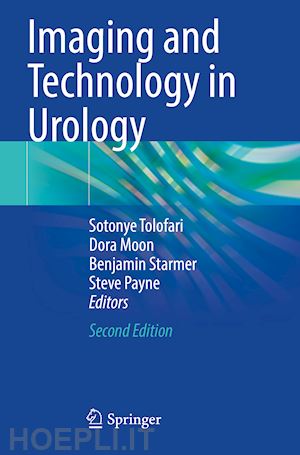
This book offers a new edition of the hugely successful title, Imaging & Technology in Urology--Principles and Clinical Applications edited by Steve Payne, Ian Eardley, Kieran O'Flynn in 2012. Essential reading for preparation of exit exams in Urology, it is used worldwide by exam candidates. Fully updated in essential areas of the book following on from recent developments in the last decade, it helps give preparation to candidates.
Part 1: Imaging Radiology.- Ch 1: Principles of X-ray production and radiation protection.- Ch 2: How to perform a clinical radiograph and use a C-arm.- Ch 3: Contrast agents.- Ch 4: Dual Energy X ray absorptiometry (DXA).- Ch 5: The physics of ultrasound and Doppler.- Ch 6: How to ultrasound a suspected renal mass.- Ch 7: How to ultrasound a painful testicle and mass.- Ch 8: How to do a trans-perineal ultrasound guided biopsy of the prostate.- Ch 9: How to manage an infected obstructed kidney.- Ch 10: Principles of computed tomography (CT).- Ch 11: How to do a CT urogram (CTU).- Ch 12: How to do a renal and adrenal CT.- Ch 13: How to do a CT in a patient with presumed upper tract trauma.- Ch 14: Principles of magnetic resonance imaging (MRI).- Ch 15: Safety in MR scanning.- Ch 16: Urological applications of MR scanning.- Ch 17: Vascular embolization techniques in urology.- Part 2: Imaging Nuclear Medicine.- Ch 18: Radionuclides and their uses in urology.- Ch 19: Counting and imaging in nuclear medicine.- Ch 20: Principles of Positron Emission Tomography (PET) scanning.- Ch 21: PET-CT imaging in prostate cancer.- Ch 22: Understanding the renogram – how it's done and how to interpret it.- Ch 23: The diuresis renogram – how it's done and how to interpret it.- Ch 24: Understanding the DMSA scan – how it's done and how to interpret it.- Ch 25: How to do a radioisotope glomerular filtration rate study.- Ch 26: Understanding the radionuclide bone scan – how it's done and how to interpret it.- Ch 27: Renography of the transplanted kidney – how it's done and how to interpret it.- Ch 28: Dynamic sentinel lymph node biopsy in penile cancer.- Part 3: Technology Diagnostic technology.- Ch 29: Urinalysis.- Ch 30: Principles of urine microscopy and micro-biological culture.- Ch 31: Urinary flow cytometry.- Ch 32: Urine cytology.- Ch 33: Histopathologial processing, staining and immuno-histochemistry.- Ch 34: Tumour markers.- Ch 35: Measurement of glomerular filtration rate (GFR).- Ch 36: Assessment of urinary tract stones.- Ch 37: Principles of pressure measurement.- Ch 38: Principles of measurement of urinary flow.- Ch 39: How to carry out a videocystometrogram (VCMG) in an adult.- Ch 40: Sphincter electromyography (EMG).- Part 4 Technology Operative.- Ch 41: Operating theatre safety.- Ch 42: Principles of decontamination.- Ch 43: Patient safety in the operating theatre environment.- Ch 44: Venous thromboembolic prevention.- Ch 45: Anticoagulants and their reversal.- Ch 46: Haemostatic agents, tissue sealants and adhesives.- Ch 47: Transfusion in urology.- Ch 48: Cell salvage in urological surgery.- Ch 49: Principles of urological endoscopes.- Ch 50: Rigid endoscope design.- Ch 51: Light Sources, light leads and camera systems.- Ch 52: Peripherals for endoscopic use.- Ch 53: Peripherals for laparoscopic use.- Ch 54: Peripherals for mechanical stone manipulation.- Ch 55: Sutures, staples and clips.- Ch 56: Contact lithotripters.- Ch 57: Monopolar diathermy.- Ch 58: Bipolar diathermy.- Ch 59: Alternatives to electro-surgery.- Ch 60: Operative tissue destruction.- Ch 61: Endoscopic use of laser energy.- Ch 62: Double J stents and nephrostomy.- Ch 63: Urinary catheters, design and usage.- Ch 64: Urological prosthetics.- Ch 65: Mesh in urological surgery.- Ch 66: Irrigant fluids and their hazards.- Ch 67: Insufflants and their hazards.- Ch 68: Laparoscopic ports.- Ch 69: Principles of robotic surgery.- Ch 70: Setting up robotic surgery.- Ch 71: Principles of tissue transfer for urologists. Part 5: Technology Interventional.- Ch 72: Neuromodulation by scaral nerve stimulation.- Ch 73: Principles of extracorporeal lithotripsy (ESWL).- Ch 74: How to carry out shockwave lithotripsy.- Ch 75: New technologies for BPH.- Ch 76: Ablative therapies.- Ch 77: Principles of radiotherapy.- Ch 78: Alternative radiotherapy techniques.- Ch 79: Augmented intravesical drug administration.- Part 6: Technology of renal failure.- Ch 80: Principles of renal replacement therapy (RRT).- Ch 81: Principles of peritoneal dialysis.- Ch 82: Haemodialysis.- Ch 83: Principles of renal transplantation.- Part 7: Assessment of technology.- Ch 84: Key concepts in the design of randomised controlled trials.- Ch 85: Reporting and Interpreting data from RCTs.- Ch 86: Health technology assessment (HTA).- Backmatter: Appendices.
Mrs Dora Moon, MBChB FRCS(Urol), Consultant Urologist
Dora Moon studied Medicine at the University of Manchester, UK graduating in 2008. She developed a interest in basic science research, further developing this whilst working as a research associate at The University of Vermont College of Medicine in the USA.
Her post-graduate surgical training was based in the North West of England, during which she sat as treasurer and committee member on the national urology trainee committee BSoT for 3 years. Dora works as an NHS consultant in Lancashire, offering a tertiary robotic surgery service for the treatment of renal cell and upper urothelial tract cancer, for patients in Lancashire and South Cumbria.
Benjamin Starmer, MBChB FRCS(Urol), Consultant UrologistSteve Payne MB MS FRCS FEBUrol, Retired Consultant Urologist
Steve qualified from the Royal Free Hospital in 1977 and was appointed Consultant Urologist at Manchester Royal Infirmary in 1988. He progressed specialist clinical interests in complex andrology and reconstructive urology at regional and national levels, whilst also having leading roles in developments in under- and post-graduate medical education. He chaired the Joint Committee on Intercollegiate Exams for the FRCS in Urology and, subsequently, the International JSCFE exam which he continues to quality assure. Steve has officiated across a number of other higher-qualification urological boards at both national and international levels and is widely published. Since retirement from clinical practice he has maintained an involvement with the production of multimedia educative material and the provision of simulation training at junior and advanced levels. In addition, Steve works with the Surgeon Wellbeing unit in the Department of Psychology at Bournemouth University, with a research interest in the peri-retirement population, and helps hands-on development of reconstructive skills for surgeons in lowand low-middle income countries in sub-Saharan Africa with colleagues from the Urolink charity.


Il sito utilizza cookie ed altri strumenti di tracciamento che raccolgono informazioni dal dispositivo dell’utente. Oltre ai cookie tecnici ed analitici aggregati, strettamente necessari per il funzionamento di questo sito web, previo consenso dell’utente possono essere installati cookie di profilazione e marketing e cookie dei social media. Cliccando su “Accetto tutti i cookie” saranno attivate tutte le categorie di cookie. Per accettare solo deterninate categorie di cookie, cliccare invece su “Impostazioni cookie”. Chiudendo il banner o continuando a navigare saranno installati solo cookie tecnici. Per maggiori dettagli, consultare la Cookie Policy.