Questo prodotto usufruisce delle SPEDIZIONI GRATIS
selezionando l'opzione Corriere Veloce in fase di ordine.
Pagabile anche con Carta della cultura giovani e del merito, 18App Bonus Cultura e Carta del Docente
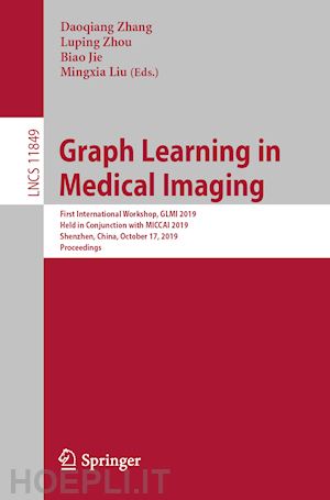
This book constitutes the refereed proceedings of the First International Workshop on Graph Learning in Medical Imaging, GLMI 2019, held in conjunction with MICCAI 2019 in Shenzhen, China, in October 2019.
The 21 full papers presented were carefully reviewed and selected from 42 submissions. The papers focus on major trends and challenges of graph learning in medical imaging and present original work aimed to identify new cutting-edge techniques and their applications in medical imaging.Graph Hyperalignment for Multi-Subject fMRI Functional Alignment.- Interactive 3D Segmentation Editing and Refinement via Gated Graph Neural Networks.- Adaptive Thresholding of Functional Connectivity Networks for fMRI-based Brain Disease Analysis.- Graph-kernel-based Multi-task Structured Feature Selection on Multi-level Functional Connectivity Networks for Brain Disease Classification.- Linking convolutional neural networks with graph convolutional networks: application in pulmonary artery-vein separation.- Comparative Analysis of Magnetic Resonance Fingerprinting Dictionaries via Dimensionality Reduction.- Learning Deformable Point Set Registration with Regularized Dynamic Graph CNNs for Large Lung Motion in COPD Patients.- Graph Convolutional Networks for Coronary Artery Segmentation in Cardiac CT Angiography.- Triplet Graph Convolutional Network forMulti-scale Analysis of Functional Connectivityusing Functional MRI.- Multi-Scale Graph Convolutional Network for Mild Cognitive Impairment Detection.- DeepBundle: Fiber Bundle Parcellation With Graph CNNs.- Identification of Functional Connectivity Features in Depression Subtypes Using a Data-Driven Approach.- Movie-watching fMRI Reveals Inter-subject Synchrony Alteration in Functional Brain Activity in ADHD.- Weakly- and Semi- Supervised Graph CNN for identifying Basal Cell Carcinoma on Pathological images.- Geometric Brain Surface Network For Brain Cortical Parcellation.- Automatic Detection of Craniomaxillofacial Anatomical Landmarks on CBCT Images using 3D Mask R-CNN.- Discriminative-Region-Aware Residual Network for Adolescent Brain Structure and Cognitive Development Analysis.- Graph Modeling for Identifying Breast Tumor Located in Dense Background of a Mammogram.- OCD Diagnosis via Smoothing Sparse Network and Stacked Sparse Auto-Encoder Learning.- A Longitudinal MRI Study of Amygdala and Hippocampal Subfields for Infants with Risk of Autism.- CNS: CycleGAN-assisted Neonatal Segmentation Model for Cross-Datasets.


Il sito utilizza cookie ed altri strumenti di tracciamento che raccolgono informazioni dal dispositivo dell’utente. Oltre ai cookie tecnici ed analitici aggregati, strettamente necessari per il funzionamento di questo sito web, previo consenso dell’utente possono essere installati cookie di profilazione e marketing e cookie dei social media. Cliccando su “Accetto tutti i cookie” saranno attivate tutte le categorie di cookie. Per accettare solo deterninate categorie di cookie, cliccare invece su “Impostazioni cookie”. Chiudendo il banner o continuando a navigare saranno installati solo cookie tecnici. Per maggiori dettagli, consultare la Cookie Policy.