libri scuola books Fumetti ebook dvd top ten sconti 0 Carrello


Torna Indietro

stoyanov danail (curatore); belagiannis vasileios (curatore); nascimento jacinto c. (curatore); lu zhi (curatore); conjeti sailesh (curatore); moradi mehdi (curatore); greenspan hayit (curatore); madabhushi anant (curatore); taylor zeike (curatore) - deep learning in medical image analysis and multimodal learning for clinical decision support

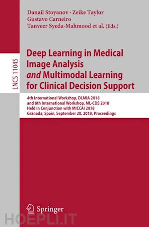
Deep Learning in Medical Image Analysis and Multimodal Learning for Clinical Decision Support 4th International Workshop, DLMIA 2018, and 8th International Workshop, ML-CDS 2018, Held in Conjunction with MICCAI 2018, Granada, Spain, September 20, 2018, Proceedings

; ; ; ; ; ; ; ;


Disponibilità: Normalmente disponibile in 15 giorni


PREZZO
54,98 €
NICEPRICE
52,23 €
SCONTO
5%


Questo prodotto usufruisce delle SPEDIZIONI GRATIS
selezionando l'opzione Corriere Veloce in fase di ordine.


Pagabile anche con Carta della cultura giovani e del merito, 18App Bonus Cultura e Carta del Docente


Facebook Twitter Aggiungi commento


Spese Gratis

Dettagli

Genere:Libro
Lingua: Inglese
Editore:

Springer

Pubblicazione: 09/2018
Edizione: 1st ed. 2018


Trama

This book constitutes the refereed joint proceedings of the 4th International Workshop on Deep Learning in Medical Image Analysis, DLMIA 2018, and the 8th International Workshop on Multimodal Learning for Clinical Decision Support, ML-CDS 2018, held in conjunction with the 21st International Conference on Medical Imaging and Computer-Assisted Intervention, MICCAI 2018, in Granada, Spain, in September 2018.

The 39 full papers presented at DLMIA 2018 and the 4 full papers presented at ML-CDS 2018 were carefully reviewed and selected from 85 submissions to DLMIA and 6 submissions to ML-CDS. The DLMIA papers focus on the design and use of deep learning methods in medical imaging. The ML-CDS papers discuss new techniques of multimodal mining/retrieval and their use in clinical decision support.


Sommario

Semi-Automated Extraction of Crohns Disease MR Imaging Markers using a 3D Residual CNN with Distance Prior.- Weakly Supervised Localisation for Fetal Ultrasound Images.- Learning to Decode 7T-like MR Image Reconstruction from 3T MR Images.- Segmentation of Head and Neck Organs-At-Risk in Longitudinal CT Scans Combining Deformable Registrations and Convolutional Neural Networks.- Iterative Segmentation from Limited Training Data: Applications to Congenital Heart Disease.- Contextual Additive Networks to Efficiently Boost 3D Image Segmentations.- Longitudinal detection of radiological abnormalities with time-modulated LSTM.- SCAN: Structure Correcting Adversarial Network for Organ Segmentation in Chest X-rays.- Active Learning for Segmentation by Optimizing Content Information for Maximal Entropy.- Rapid Training Data Generation for Tissue Segmentation Using Global Approximate Block-Matching with Self-Organizing Maps.- Reinforced Auto-Zoom Net: Towards Accurate and Fast Breast Cancer Segmentation in Whole-slide Images.- Deep semi-supervised segmentation with weight-averaged consistency targets.- Focal Dice Loss and Image Dilation for Brain Tumor Segmentation.- Automatic Detection of Patients with a High Risk of Systolic Cardiac Failure in Echocardiography.- Unsupervised feature learning for outlier detection with stacked convolutional autoencoders, siamese networks and Wasserstein autoencoders: application to epilepsy detection.- Automatic myocardial strain imaging in echocardiography using deep learning.- 3D Convolutional Neural Networks for Classification of Functional Connectomes.- Computed Tomography Image Enhancement using 3D Convolutional Neural Network.- Deep Particle Tracker: Automatic Tracking of Particles in Fluorescence Microscopy Images Using Deep Learning.- A Unified Framework Integrating Recurrent Fully-convolutional Networks and Optical Flow for Segmentation of the Left Ventricle in Echocardiography Data.- Learning Optimal Deep Projection of 18 F-FDG PET Imaging for Early Differential Diagnosis of Parkinsonian Syndromes.- Learning to Segment Medical Images with Scribble-Supervision Alone.- Unsupervised Probabilistic Deformation Modeling for Robust Diffeomorphic Registration.- TreeNet: Multi-Loss Deep Learning Network to Predict Branch Direction for Extracting 3D Anatomical Trees.- Active Deep Learning with Fisher Information for Patch-wise Semantic Segmentation.- UOLO - automatic object detection and segmentation in biomedical images.- Pediatric Bone Age Assessment Using Deep Convolutional Neural Networks.- Multi-Scale Residual Network with Two Channels of Raw CT Image and Its Differential Excitation Component for Emphysema Classification.- Nonlinear adaptively learned optimization for object localization in 3D medical images.- Automatic Segmentation of Pulmonary Lobes Using a Progressive Dense V-Network.- UNet++: A Nested U-Net Architecture for Medical Image Segmentation.- MTMR-Net: Multi-Task Deep Learning with Margin Ranking Lossfor Lung Nodule Analysis.- PIMMS: Permutation Invariant Multi-Modal Segmentation.- Handling Missing Annotations for Semantic Segmentation with Deep ConvNets.- 3D Deep Affine-Invariant Shape Learning for Brain MR Image Segmentation.- ScarGAN: Chained Generative Adversarial Networks to Simulate Pathological Tissue on Cardiovascular MR Scans.- Unpaired Deep Cross-modality Synthesis with Fast Training .- Monte-Carlo Sampling applied to Multiple Instance Learning for Histological Image Classification.- Unpaired Brain MR-to-CT Synthesis using a Structure-Constrained CycleGAN.- A Multi-Scale Multiple Sclerosis Lesion Change Detection in a Multi-Sequence MRI.- Multi-task Sparse Low-rank Learning for Multi-classification of Parkinson’s Disease.- Optic Disc segmentation in Retinal Fundus Images using Fully Convolutional Network and Removal of False-positives Based on Shape Features.- Integrating deformable modeling with 3D deep neural network segmentation.


Altre Informazioni

ISBN:

9783030008888

Condizione: Nuovo
Collana: Lecture Notes in Computer Science
Dimensioni: 235 x 155 mm
Formato: Brossura
Illustration Notes:XVII, 387 p. 197 illus., 149 illus. in color.
Pagine Arabe: 387
Pagine Romane: xvii


Dicono di noi